- About Us
- Ilizarov Technique
- Process
- Case Studies
Fractures
• Acute Fracture - Comminuted Fracture Distal Humerus - Compound Fracture & VAC • Nonunion - Proximal Tibia - Tibia - Distal Humerus • Malunion - Femur Bone Deformity
• Acute Correction
- LRS - Bilateral Tibia - LRS - Rotational Deformity Femur(with Shortening) - LRS - Oblique Plane Deformity Femur - Nailing • Ilizarov Correction - Proximal Tibia - Distal Femur • Hexapod Correction - Metaphyseal Dysplasia Bone Infection
• Infection - Femur • Infected Nonunion - Proximal Femur Joint Problems (Arthritis)
• Hip Replacement • Knee Replacement • Intra Articular Osteotomy • High Tibial Osteotomy (HTO) • Arthrolysis - Elbow • Ilizarov Hip Reconstruction - FAQs
- Testimonials
News & Updates


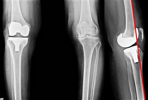
Knee Replacement
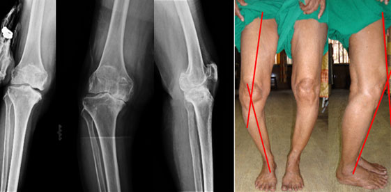
| 70 year old lady, presents with pain in both the knees right more than left, difficulty in walking, bowing of both legs. The x-rays show osteoarthritis of knee joint, destruction of joint cartilage, varus deformity. The pictures on the right show the clinical appearance. When looking from the front there was severe bowing of the legs and on looking from the side there was flexion deformity (knees bent). Both the deformities adding mechanical disadvantage to the destroyed joint cartilage. | ||
 |
||
|
||
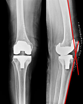
| 3 months post surgery | ||
The appearance 3 months after surgery. The deformities are resolved, compare with the un-operated side . The range of motion is good and more importantly there is no pain. |
||
 |
||
| 3 months post surgery | ||
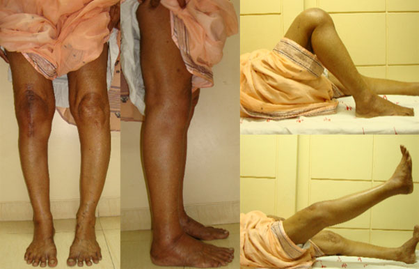
The x-rays after 3 months of surgery. The flexion deformity has resolved. The un-operated side also shows damage, but the patient is not symptomatic on that side so no need for surgery till such time. |
||
 |
||